Home
/ Diagram Rib Cage With Organs : Image Photos A699bef7 3c52 46ef A515 5716c4f56c261316411967947 Thumb For Term Side Of Card Human Rib Cage Rib Cage Anatomy Human Ribs _ The top edge of the manubrium has a depression called the suprasternal or jugular notch.
Diagram Rib Cage With Organs : Image Photos A699bef7 3c52 46ef A515 5716c4f56c261316411967947 Thumb For Term Side Of Card Human Rib Cage Rib Cage Anatomy Human Ribs _ The top edge of the manubrium has a depression called the suprasternal or jugular notch.
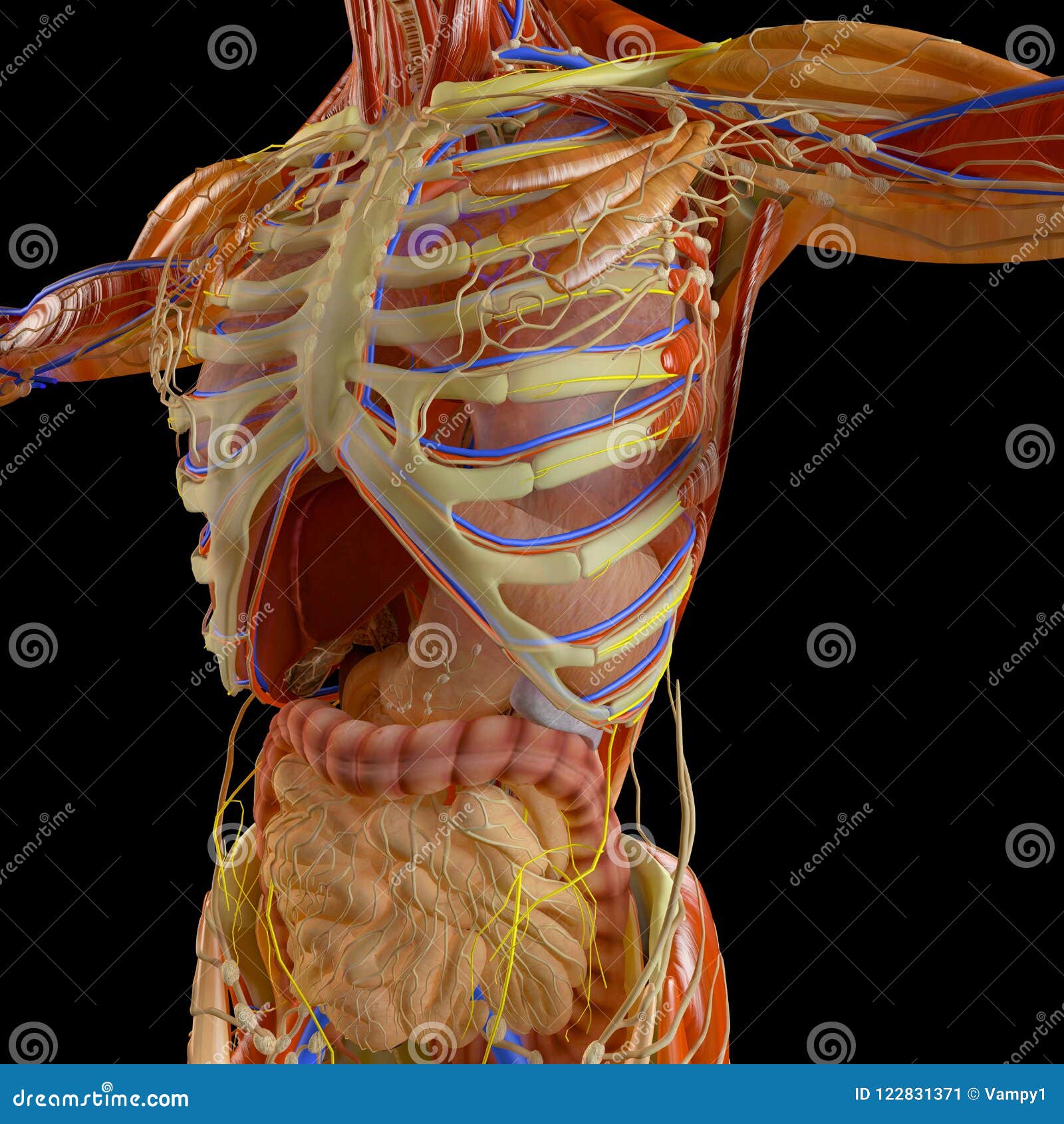
Diagram Rib Cage With Organs : Image Photos A699bef7 3c52 46ef A515 5716c4f56c261316411967947 Thumb For Term Side Of Card Human Rib Cage Rib Cage Anatomy Human Ribs _ The top edge of the manubrium has a depression called the suprasternal or jugular notch.. You have two kidneys located below your rib cage. Diagram of human body, liver rib cage, rib cage diagram labeled, rib cage diagram numbered, rib cage diaphragm, rib cage heart, rib cage organs anatomy, rib cage pain, stomach, diagram of human body, liver rib cage, rib cage diagram labeled, rib cage diagram numbered, rib cage diaphragm, rib cage. The rib cage protects vital internal organs. Posted on december 22, 2018december 22, 2018. Anatomy of the rib cage diagram.
Rib cage anatomy the rib cage, shaped in a mild cone shape and more flexible than most bone sets, is made up of varying elements such as the thoracic vertebra, 12 equally paired ribs, costal cartilage, and held together anteriorly by the sternum. In this video we discuss the structure of the rib cage or thoracic cage. Other female reproductive structures serve as. Our latest youtube film is ready to run. They also have a role in ventilation;
Ribcage Anatomy Stock Illustrations 591 Ribcage Anatomy Stock Illustrations Vectors Clipart Dreamstime from thumbs.dreamstime.com Other female reproductive structures serve as. They also have a role in ventilation; The ribs are curved, flat bones which form the majority of the thoracic cage.they are extremely light, but highly resilient; Of all human body skeleton system upper limbs anatomy. Rib cage, in vertebrate anatomy, basketlike skeletal structure that forms the chest, or thorax, and is made up of the ribs and their corresponding attachments to the sternum (breastbone) and the vertebral column.the rib cage surrounds the lungs and the heart, serving as an important means of bony protection for these vital organs.in total, the rib cage consists of the 12 thoracic vertebrae and. We cover the different bones that make up the rib cage and some of the functions of. These organ systems interact to produce coordinated, active, healthy and intelligent human body. Each are symmetrically paired on a right and left side.
Anatomy of the rib cage diagram.
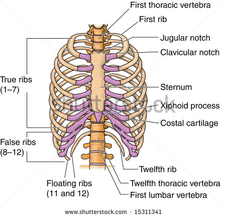
Anatomy of the elbow 12 photos of the anatomy of the elbow anatomy and biomechanics of the elbow pdf, anatomy elbow fossa, anatomy of elbow dislocation, anatomy of the elbow bones, anatomy of the elbow muscles, human anatomy, anatomy and biomechanics of the elbow pdf, anatomy elbow fossa, anatomy of elbow dislocation. The sternum, commonly known as the breastbone, is a long, narrow flat bone that serves as the keystone of the rib cage and stabilizes the thoracic skeleton. The area just under the bottom part of the rib cage is the abdomen and not the chest cavity as is commonly thought. The rib cage protects vital organs, such as the heart and lungs. An injury to the left rib cage can give rise to severe pain in under the ribs, particularly when the affected person is breathing deeply. How to draw human spine and rib cage diagram. The sternum is a flat bone that is made up of three parts, the (1) manubrium, (2) body, and the (3) xiphoid process. Anatomy of the rib cage diagram. The last diagram shows how the ribs are connected to the vertebral column or spine. Related posts of abdominal diagram with ribs anatomy of the elbow. The primary responsibilities of the ribcage involve protecting the thoracic visceral organs, enclosing the thoracic visceral organs, and is included. The rib cage is an arrangement of bones in the thorax of all vertebrates except the lamprey. There are 12 pairs of ribs and they attach to the spine.
2006 kia optima belt diagram. They also have a role in ventilation; Of all human body skeleton system upper limbs anatomy. For more anatomy content please follow us and visit our website: It encloses and protects the heart and lungs.
Bones Of The Human Chest Rib Cage Stock Vector Colourbox from www.colourbox.com Anatomy of the elbow 12 photos of the anatomy of the elbow anatomy and biomechanics of the elbow pdf, anatomy elbow fossa, anatomy of elbow dislocation, anatomy of the elbow bones, anatomy of the elbow muscles, human anatomy, anatomy and biomechanics of the elbow pdf, anatomy elbow fossa, anatomy of elbow dislocation. 2006 kia optima belt diagram. The rib cage is collectively made up of long, curved individual. In this video we discuss the structure of the rib cage or thoracic cage. The rib cage protects vital internal organs. The majority of liver mass is in the upper right side of the abdomen, just under the rib cage. Humans have five vital organs that are essential for survival. Of all human body skeleton system upper limbs anatomy.
An injury to the left rib cage can give rise to severe pain in under the ribs, particularly when the affected person is breathing deeply.
Rib cage anatomy rib cage diagram with organs anatomy of rib. What organ is under your right rib cage. Male body internal organs chart with labels on white background. Related posts of abdominal diagram with ribs anatomy of the elbow. Rib cage diagram with organs. The sternum, commonly known as the breastbone, is a long, narrow flat bone that serves as the keystone of the rib cage and stabilizes the thoracic skeleton. You have two kidneys located below your rib cage. The ribs partially enclose and protect the chest cavity, where many vital organs (including the heart and the lungs) are located. Diagram of human body, liver rib cage, rib cage diagram labeled, rib cage diagram numbered, rib cage diaphragm, rib cage heart, rib cage organs anatomy, rib cage pain, stomach, diagram of human body, liver rib cage, rib cage diagram labeled, rib cage diagram numbered, rib cage diaphragm, rib cage. We cover the different bones that make up the rib cage and some of the functions of. Our latest youtube film is ready to run. The following diagrams are the rib cage diagrams. Rib cage illustration stock photos rib cage.
The rib cage protects vital organs, such as the heart and lungs. In this image, you will find thoracic vertebrum, costochondral joint, costal cartilage, costal margin, costal arch, thoracic vertebrum, xiphoid process, xiphisternal joint, body, manubrial sternal joint, manubrium, the sternal notch in it. Posted on december 22, 2018december 22, 2018. These are the brain, heart, kidneys, liver and lungs. Related posts of rib cage diagram with organs anatomy of human stomach.
Besides The Ribs Which Bones Protect The Lungs And The Heart Socratic from useruploads.socratic.org The sternum is a flat bone that is made up of three parts, the (1) manubrium, (2) body, and the (3) xiphoid process. Contributing to their role in protecting the internal thoracic organs. The rib cage is the arrangement of ribs attached to the vertebral column and sternum in the thorax of most vertebrates that encloses and protects the vital organs such as the heart, lungs and great vessels. Related posts of rib cage diagram with organs anatomy of human stomach. 05.11.2019 · rib cage diagram with organs, find out more about rib cage diagram with organs. Rib cage, in vertebrate anatomy, basketlike skeletal structure that forms the chest, or thorax, and is made up of the ribs and their corresponding attachments to the sternum (breastbone) and the vertebral column.the rib cage surrounds the lungs and the heart, serving as an important means of bony protection for these vital organs.in total, the rib cage consists of the 12 thoracic vertebrae and. The rib cage is collectively made up of long, curved individual. Introduction to the structure of the ribcage and ribs:
Each are symmetrically paired on a right and left side.
Introduction to the structure of the ribcage and ribs: The top edge of the manubrium has a depression called the suprasternal or jugular notch. The area just under the bottom part of the rib cage is the abdomen and not the chest cavity as is commonly thought. Related posts of abdominal diagram with ribs anatomy of the elbow. How to draw human spine and rib cage diagram. Several muscles that move the arms, head, and neck have their origins on the sternum. Rib cage human anatomy organs. The human rib cage is made up of 12 paired rib bones; These are the brain, heart, kidneys, liver and lungs. Rib cage anatomy rib cage diagram with organs anatomy of rib. The primary responsibilities of the ribcage involve protecting the thoracic visceral organs, enclosing the thoracic visceral organs, and is included. The rib cage protects vital internal organs. 3d illustration of human body skeleton system upper limbs anatomy
